
Academic Papers
Guidelines for the Academic Paper Contest for 2026
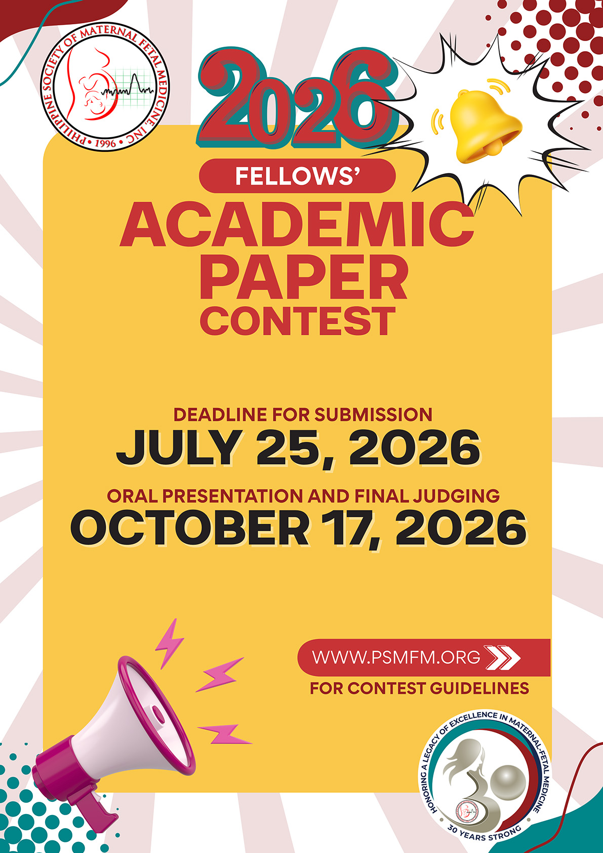

Guidelines for the Academic Paper Contest for 2026
LEARN MORE
PSMFM Contest Winners


Philippine Society of Maternal Fetal Medicine
2024 Research Paper Contest First Place
October 19, 2024
Applied deep learning networks in the creation of a risk assessment tool to predict spontaneous preterm birth using maternal risk factors, cervical length and hardness ratio, and phosphorylated insulin-like growth factor binding protein-1
Primary Author: Grace Lynn S. Estanislao, MD
Co-Authors: Prof. Rafael Alampay, Maria Rosario C. Cheng, MD and Zarinah G. Gonzaga, MD
The Medical City
Abstract
Predicting spontaneous preterm birth (SPTB) and its occurrence within 48 hours to 7 days is desirable for optimizing maternal and neonatal outcomes. Most existing prediction models lack clinical applicability, underscoring the need for improved prediction tools. The main objective of this study was to apply deep learning to generate prediction models that combine maternal risk factors, cervical length (CL) and hardness ratio (HR) measurements, and phosphorylated insulin- like growth factor binding protein-1 (phIGFBP-1) results to predict SPTB in pregnant women with singleton gestation from 24 to 35 6/7 weeks age of gestation. Specific objectives included: (1) creating multiple prediction models and selecting the most suitable model, (2) training it to predict the risk of preterm delivery, delivery within 7 days, and delivery within 48 hours, (3) testing the models, and (4) embedding them in a mobile application to generate individualized risk scores. Data on maternal age, obstetric history, CL, HR, and phIGFBP-1 testing were collected from 887 records from August 2018 to December 2023. Multiple deep learning models were trained, validated and evaluated for predicting SPTB and delivery within specific time frames. The CL-only model for predicting preterm delivery outperformed other models (F1 score 84.21%, AUC 94%, sensitivity 80%, specificity 90%, PPV 85.35%, NPV 81.82%), with a recommended 35% threshold for intervention. A variation of this model for predicting delivery within 48 hours also had good performance (F1 score 76.19%, AUC 83%, sensitivity 80%, specificity 70%, PPV 75.25%, NPV 77.78%). AUCs of these CL models were notably higher than existing models in literature. Remarkably, the addition of more predictors (i.e., HR, phIGFBP-1) led to a decline in the performance of the prediction models. The developed mobile application provides individualized risk scores for SPTB, offering a user-friendly adjunctive tool for clinicians to support decision-making in managing preterm labor.
Keywords: artificial intelligence; cervical elastography; cervical hardness ratio; cervical length; deep learning; phIGFBP-1; phosphorylated insulin-like growth factor binding protein-1; prediction tool; prediction model; prediction tool; predictive model; preterm labor; risk assessment; spontaneous preterm birth

Philippine Society of Maternal Fetal Medicine
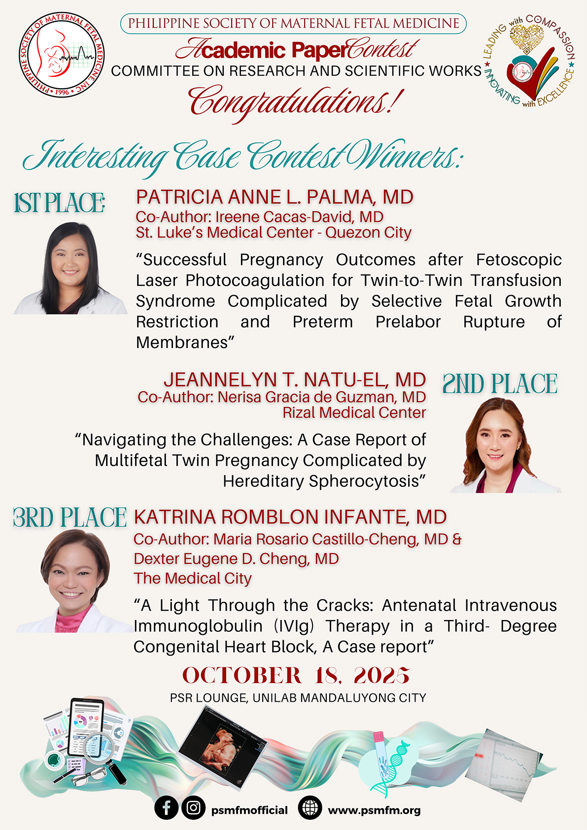
2023 Interesting Case Contest Winners
November 6, 2023
First Place Winner
SUCCESFUL PREGNANCY IN A PATIENT WITH MULTIPLE SEVERE ORGAN MANIFESTATION OF SYSTEMIC LUPUS ERYTHEMATOSUS
Primary author: Laura Andrea B. Rivera-Mapili, MD, FPOGS*
Fellow, Section of Maternal Fetal Medicine, Department of Obstetrics and Gynecology, Quirino Memorial Medical Center
Co-author: Maria Czarina V. Mendoza, MD FPOGS FPSMFM
Head, Section of Maternal Fetal Medicine, Department of Obstetrics and Gynecology, Quirino Memorial Medical Center
*Corresponding author: labriveramd@gmail.com
ABSTRACT
Systemic lupus erythematosus (SLE) is a heterogenous, highly complex autoimmune disease which involves various organ systems with diverse manifestations. The incidence is slightly higher in women with reproductive age, hence special interest has been given to the interplay between the disease behavior and pregnancy. This is a case of a 24-year-old primigravid who was diagnosed with SLE with neuropsychiatric manifestation five years prior to pregnancy. The clinical course within two years of diagnosis was exceptionally complicated despite disease quiescence of more than 6 months prior to pregnancy, making her a clinical enigma. Patient had multiple severe organ manifestations including nephritis, mastitis and severe anemia during pregnancy which necessitated multiple glucocorticoid therapy. The treatment needed to control the life- threatening disease flares led to development of more maternal and fetal problems such as worsening hypertension, hyperglycemia, fetal growth restriction and preterm labor. A multidisciplinary approach to the management of this case, close multi-organ surveillance and thorough maternal-fetal monitoring led to a successful pregnancy outcome. Elucidating the mechanism by which her history of flares affected her pregnancy course also aided in crafting a comprehensive treatment plan for a favorable outcome.
Keywords: SLE, systemic lupus erythematosus, flare, adverse pregnancy outcome
Second Place Winner
CHORIOAMNIOTIC MEMBRANE SEPARATION AFTER AMNIOREDUCTION: A CASE REPORT
Primary author: Sheryl Lyn M. Pepito, MD*
Fellow, Division of Maternal-Fetal Medicine, Department of Obstetrics and
Gynecology, University of the Philippines-Philippine General Hospital, Manila
Co-author: Joselito A. Santiago, MD, FPOGS, FPSMFM, FPSUOG
Division Chief, Maternal-Fetal Medicine, Department of Obstetrics and
Gynecology, University of the Philippines-Philippine General Hospital, Manila
*Corresponding author: smpepito1@up.edu.ph
ABSTRACT
We present a 40-year-old secundigravida who had threatened preterm labor and sonologic findings of a small gastric bubble, fetal growth restriction, polyhydramnios, rockerbottom feet, clenched fists and overlapping fingers, suggestive of fetal aneuploidy. She underwent amnioreduction, both as a therapeutic and diagnostic procedure. Chorioamniotic membrane separation was detected sonographically two weeks post amnioreduction and subsequently thereafter. Weekly biophysical profile and doppler studies were done until admission for labor induction at 37 weeks gestation. During which time, the official karyotyping showed trisomy 18, consistent with her neonatal karyotyping. Primary cesarean section was performed for recurrent late decelerations. Grossly, the amniotic membrane was completely separated from the chorionic membrane all around the placenta and was only attached at the umbilical cord insertion. Despite the lack of guidelines on the antenatal surveillance of chorioamniotic membrane separation, we were able to deliver a live fetus at term.
Keywords: amnioreduction, aneuploidy, chorioamnionitic membrane separation
Third Place Winner
SOLID PSEUDOPAPILLARY NEOPLASM OF THE PANCREAS DURING PREGNANCY PRESENTING AS GASTROINTESTINAL STROMAL TUMOR: A CASE REPORT AND REVIEW OF LITERATURE
Primary author: Stephanie S. Causin, MD, FPOGS*
Fellow, Section of Maternal Fetal Medicine, Institute for Women’s Health
The Medical City, Ortigas Avenue, Pasig City
Co-author: Zarinah G. Gonzaga, MD, FPOGS, FPSMFM, FPSUOG
Consultant, Section of Maternal Fetal Medicine, Institute for Women’s Health
The Medical City, Ortigas Avenue, Pasig City
*Corresponding author: stephaniecausin@gmail.com
ABSTRACT
Solid pseudopapillary neoplasm (SPN) is a rare tumor that can complicate pregnancy. More than its rarity, SPNs are unique neoplasms because of their obscure histogenesis, cytology, immunohistochemical profile, and imaging characteristics. This report describes a case of a 32-year-old gravida 2 para 1 (1001) seen at 24 weeks with an intraabdominal mass. The patient presented with a long-standing history of abdominal mass with the working impression of gastrointestinal stromal tumor. We employed a multidisciplinary approach to closely monitor tumor growth, ensure maternal and fetal well-being, avert complications, and avoid unnecessary clinical interventions. Histopathologic evaluation and immunohistochemistry studies of representative specimens taken at the time of delivery revealed the diagnosis of SPN of the pancreas. At the time of writing, the patient is awaiting schedule for definitive surgery. To the best of our knowledge, this is the first documented case of SPN complicating pregnancy in the Philippines.
Keywords: Intra-abdominal Mass, Pregnancy, Solid Pseudopapillary Neoplasm (SPN)

Philippine Society of Maternal Fetal Medicine
2023 Research Contest Winner
November 6, 2023
A SONOGRAPHIC EVALUATION ON AGREEMENT AND TIME EFFICIENCY OF FETAL CENTRAL NERVOUS SYSTEM BIOMETRY USING SEMI-AUTOMATED FIVE- DIMENSIONAL (5D) ULTRASOUND VERSUS STANDARD TWO-DIMENSIONAL (2D) ULTRASOUND IN A PHILIPPINE TERTIARY HOSPITAL
Primary author: Lizzette R. Caro-Alquiros, MD, MBA, FPOGS*
Fellow, Section of Maternal Fetal Medicine, Institute for Women’s Health,
The Medical City, Ortigas Avenue, Pasig City
Co-authors: Zarinah G. Gonzaga, MD, MBA, MHM, FPOGS, FPSMFM, FPSUOG
Irene B. Quinio, MD, MBAH, FPOGS, FPSMFM, FPSUOG
Consultants, Section of Maternal Fetal Medicine, Institute for Women’s Health,
The Medical City, Ortigas Avenue, Pasig City
*Corresponding author: lizzettecaro@gmail.com
ABSTRACT
Objective: The objectives of this research are to evaluate the agreement of cranial biometric measurements and to determine if there is a significant difference in the time needed to complete the evaluation using standard 2D and semi-automated 5D ultrasound.
Methods: An analytical cross-sectional study was employed on 93 women who underwent pelvic ultrasound scans from August 2022 to October 2022 in a tertiary hospital. Basic biometric fetal CNS measurements were acquired using 2D ultrasound followed by 5D CNS ultrasound. Bland-Altman plots were used to evaluate the agreement of the measurements obtained. The difference in the time to completion was determined using independent t-test.
Results: The 5D CNS ultrasound successfully measured the basic fetal CNS biometry in 90 out of 93 (96.8%) fetuses. It showed an average of 94.4 percent agreement on all measurements. The 5D CNS ultrasound takes a shorter time of 90 s to completion in comparison to 99 s using the 2D method. However, this 9-second difference was not statistically significant (p=0.076). Upon stratification of the study population per trimester, in the second trimester, it took 76 s with 5D CNS vs 89 s with 2D, resulting to a statistically significant 13-second difference (p=0.044). In the third trimester, 5D CNS took 105 s vs 108 s with 2D. The 3-second difference was not statistically significant (p=0.614).
Conclusion: Our study found that 5D CNS ultrasound measurements showed agreement with 2D ultrasound. The time to completion of the scan using this technology is faster when used for second trimester pregnancies. However, time to completion could be affected by fetal-dependent and operator-dependent factors. Therefore, application of this new technology has the potential to improve workflow efficiency after the necessary training on 3D sonography and 5D CNS ultrasound software.
Keywords: 5D CNS; AI; fetal brain; fetal CNS; ultrasound
PSMFM Contest Winners
LEARN MORE
Philippine Journal of Obstetrics and Gynecology

Special Issue: Philippine Society of Maternal Fetal Medicine
Volume 48 – Issue 2 – April-June 2024
Available for viewing and download
- Perspective article of Dr Emerson D. Tan: State of Maternal Fetal Medicine in the Philippines
- Program Evaluation and early outcomes of a severe preeclampsia and eclampsia maternal safety bundle in a single institution in the Phils by Zarinah G. Gonzaga, Maria Rosario Castillo-Cheng, JC Macalintal, Lizzette Caro-Alquiros, Stephanie Causin, Grace Lynn S. Estanislao (original article)
- (Original article) A sonographic evaluation on agreement and time efficiency of fetal central nervous system biometry using a semi-automated five-dimensional ultrasound versus two-dimensional ultrasound in a Philippine tertiary hospital by Lizzette Caro-Alquiros, Zarinah G. Gonzaga , Irene B. Quinio
- (Original article) Association of global cardiac sphericity index and neonatal outcomes of appropriate for gestational age fetuses, small for gestational age fetuses and growth-restricted fetuses delivered at term in Dr Jose Fabella Memorial Hospital: a prospective cohort study – by Brenan Ian Capuno and Roberto Montana
- (Original article) Assessing the resident physicians’ perceptions of the use webinars to support training during the COVID 19 pandemic – by M Misuno , Valerie T. Guinto
- (Case report) Metastatic colon adenocarcinoma in pregnancy – by Deverly Rina V. reyes, Jemimah T. Cartagena-Lim, Mario Philip R Festin
- (Case report) Recurrent dedifferentiated retroperitoneal liposarcoma complicating pregnancy – by Jemimah T. Cartagena-Lim , Kristine Therese Elises-Molon
- Solid pseudopapillary neoplasm of the pancreas during pregnancy presenting as gastrointestinal stromal tumor: a case report and review of literature- by Stephanie Causin , Zarinah G. Gonzaga
Philippine Journal of Obstetrics and Gynecology
LEARN MORE
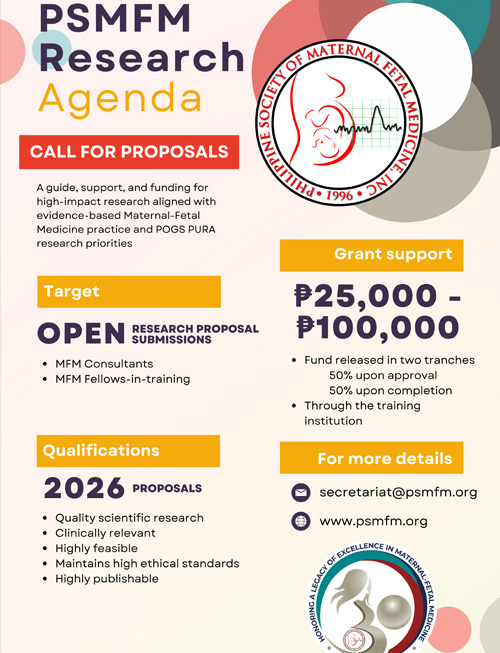
PSMFM Research Agenda
Are you a young researcher passionate about maternal fetal medicine?
Do you have a groundbreaking study that could revolutionize the field?
We invite you to submit your research for consideration as part of the Maternal-Fetal Medicine research agenda.
Why submit your research?
Gain recognition: Showcase your work to a global audience of experts in maternal fetal medicine.
Network with peers: Connect with like-minded researchers and establish valuable collaborations.
Receive feedback: Get expert insights and recommendations to enhance your research.
Contribute to the field: Help advance our understanding of maternal fetal health and improve patient outcomes.
The following are considered as research priorities:
Responsive health systems
Enhance and extend healthy lives
Holistic approach to health and wellness
Health resiliency
Global competitiveness and innovation
Research in equity and health
Selected submissions will have more opportunities to:
Present their research at the conference
Publish their findings in a peer-reviewed journal
Receive mentorship from leading experts in the field
Be awarded with funding from the society
Don’t miss this chance to share your research and make a lasting impact on maternal fetal medicine. Submit your abstract and proposals today!
For more information, please contact: PSMFM Secretariat.
Or download information sheet HERE
PSMFM Research Agenda
LEARN MORE
